Source:- Google.com.pk
Muscle & Fitness is an American fitness and bodybuilding magazine founded by Joe Weider, but now published by American Media, Inc.
Muscle & Fitness has a more mainstream fitness & bodybuilding lifestyle focus than its companion publication, Flex, which mainly covers more specialised "hardcore" and professional bodybuilding topics. It offers many exercise and nutritional tips, while at the same time advertising a variety of nutritional supplements from companies such as BSN.
Many professional bodybuilders are featured in each monthly issue of Muscle & Fitness, like Ronnie Coleman, Gustavo Badell, Darrem Charles, Sagi Kalev and Jay Cutler.
Muscle & Fitness has also featured actors, natural bodybuilders, athletes and sports personalities such as: Arnold Schwarzenegger, 50 Cent, Evander Holyfield, Joe Weider, Dwayne Johnson, Sebastian Siegel, Jean-Claude Van Damme, Emilien De Falco, Lawrence Leritz, Mike O'Hearn, and the March 2006 issue featured World Wrestling Entertainment chairman Vince McMahon and superstar Triple H. Triple H also featured in another issue at the end of 2009. It also features a number of figure competitors such as Davana Medina, Jenny Lynn, Monica Brant. And it featured on the cover of the February 2008 issue former UFC Heavyweight Champion Brock Lesnar. Muscle and Fitness placed a lot of emphasis on Wrestlers and MMA fighters towards the end of 2009 and into 2010. Editions included WWE Superstar John Cena, former WWE Superstars "Stone Cold" Steve Austin and John Morrison, and UFC fighters Todd Duffee and Nate Marquardt.Muscle is a soft tissue found in most animals. Muscle cells contain protein filaments of actin and myosin that slide past one another, producing a contraction that changes both the length and the shape of the cell. Muscles function to produce force and motion. They are primarily responsible for maintaining and changing posture, locomotion, as well as movement of internal organs, such as the contraction of the heart and the movement of food through the digestive system via peristalsis.
Muscle tissues are derived from the mesodermal layer of embryonic germ cells in a process known as myogenesis. There are three types of muscle, skeletal or striated, cardiac, and smooth. Muscle action can be classified as being either voluntary or involuntary. Cardiac and smooth muscles contract without conscious thought and are termed involuntary, while the skeletal muscles contract upon command.[citation needed] Skeletal muscles in turn can be divided into fast and slow twitch fibers.
Muscles are predominantly powered by the oxidation of fats and carbohydrates, but anaerobic chemical reactions are also used, particularly by fast twitch fibers. These chemical reactions produce adenosine triphosphate (ATP) molecules which are used to power the movement of the myosin heads.[citation needed]
The term muscle is derived from the Latin musculus meaning "little mouse" perhaps because of the shape of certain muscles or because contracting muscles look like mice moving under the skin.
The anatomy of muscles includes gross anatomy, which comprises all the muscles of an organism, and microanatomy, which comprises the structures of a single muscle.
The body contains three types of muscle tissue: (a) skeletal muscle, (b) smooth muscle, and (c) cardiac muscle. (Same magnification)Muscle tissue is a soft tissue, and is one of the four fundamental types of tissue present in animals. There are three types of muscle tissue recognized in vertebrates:
Skeletal muscle or "voluntary muscle" is anchored by tendons (or by aponeuroses at a few places) to bone and is used to effect skeletal movement such as locomotion and in maintaining posture. Though this postural control is generally maintained as an unconscious reflex, the muscles responsible react to conscious control like non-postural muscles. An average adult male is made up of 42% of skeletal muscle and an average adult female is made up of 36% (as a percentage of body mass).
Smooth muscle or "involuntary muscle" is found within the walls of organs and structures such as the esophagus, stomach, intestines, bronchi, uterus, urethra, bladder, blood vessels, and the arrector pili in the skin (in which it controls erection of body hair). Unlike skeletal muscle, smooth muscle is not under conscious control.Cardiac muscle (myocardium), is also an "involuntary muscle" but is more akin in structure to skeletal muscle, and is found only in the heart.Cardiac and skeletal muscles are "striated" in that they contain sarcomeres that are packed into highly regular arrangements of bundles; the myofibrils of smooth muscle cells are not arranged in sarcomeres and so are not striated. While the sarcomeres in skeletal muscles are arranged in regular, parallel bundles, cardiac muscle sarcomeres connect at branching, irregular angles (called intercalated discs). Striated muscle contracts and relaxes in short, intense bursts, whereas smooth muscle sustains longer or even near-permanent contractions.Skeletal (voluntary) muscle is further divided into two broad types: slow twitch and fast twitch:
Type I, slow twitch, or "red" muscle, is dense with capillaries and is rich in mitochondria and myoglobin, giving the muscle tissue its characteristic red color. It can carry more oxygen and sustain aerobic activity using fats or carbohydrates as fuel.Slow twitch fibers contract for long periods of time but with little force.Type II, fast twitch muscle, has three major subtypes (IIa, IIx, and IIb) that vary in both contractile spee and force generated.Fast twitch fibers contract quickly and powerfully but fatigue very rapidly, sustaining only short, anaerobic bursts of activity before muscle contraction becomes painful. They contribute most to muscle strength and have greater potential for increase in mass. Type IIb is anaerobic, glycolytic, "white" muscle that is least dense in mitochondria and myoglobin. In small animals (e.g., rodents) this is the major fast muscle type, explaining the pale color of their flesh.The density of mammalian skeletal muscle tissue is about 1.06 kg/liter.This can be contrasted with the density of adipose tissue (fat), which is 0.9196 kg/liter.This makes muscle tissue approximately 15% denser than fat tissue.
A chicken embryo, showing the paraxial mesoderm on both sides of the neural fold. The anterior (forward) portion has begun to form somites (labeled "primitive segments").
All muscles are derived from paraxial mesoderm.The paraxial mesoderm is divided along the embryo's length into somites, corresponding to the segmentation of the body (most obviously seen in the vertebral column.Each somite has 3 divisions, sclerotome (which forms vertebrae), dermatome (which forms skin), and myotome (which forms muscle).The myotome is divided into two sections, the epimere and hypomere, which form epaxial and hypaxial muscles, respectively.The only epaxial muscles in humans are the erector spinae and small intervertebral muscles, and are innervated by the dorsal rami of the spinal nerves. All other muscles, including those of the limbs are hypaxial, and inervated by the ventral rami of the spinal nerves
During development, myoblasts (muscle progenitor cells) either remain in the somite to form muscles associated with the vertebral column or migrate out into the body to form all other muscles.Myoblast migration is preceded by the formation of connective tissue frameworks, usually formed from the somatic lateral plate mesoderm.Myoblasts follow chemical signals to the appropriate locations, where they fuse into elongate skeletal muscle cells.
A skeletal muscle fiber is surrounded by a plasma membrane called the sarcolemma, which contains sarcoplasm, the cytoplasm of muscle cells. A muscle fiber is composed of many fibrils, which give the cell its striated appearance.Skeletal muscles are sheathed by a tough layer of connective tissue called the epimysium. The epimysium anchors muscle tissue to tendons at each end, where the epimysium becomes thicker and collagenous. It also protects muscles from friction against other muscles and bones. Within the epimysium are multiple bundles called fascicles, each of which contains 10 to 100 or more muscle fibers collectively sheathed by a perimysium. Besides surrounding each fascicle, the perimysium is a pathway for nerves and the flow of blood within the muscle.The threadlike muscle fibers are the individual muscle cells (myocytes), and each cell is encased within its own endomysium of collagen fibers Thus, the overall muscle consists of fibers (cells) that are bundled into fascicles, which are themselves grouped together to form muscles. At each level of bundling, a collagenous membrane surrounds the bundle, and these membranes support muscle function both by resisting passive stretching of the tissue and by distributing forces applied to the muscle. Scattered throughout the muscles are muscle spindles that provide sensory feedback information to the central nervous system. (This grouping structure is analogous to the organization of nerves which uses epineurium, perineurium, and endoneurium).
This same bundles-within-bundles structure is replicated within the muscle cells. Within the cells of the muscle are myofibrils, which themselves are bundles of protein filaments. The term "myofibril" should not be confused with "myofiber", which is a simply another name for a muscle cell. Myofibrils are complex strands of several kinds of protein filaments organized together into repeating units called sarcomeres. The striated appearance of both skeletal and cardiac muscle results from the regular pattern of sarcomeres within their cells. Although both of these types of muscle contain sarcomeres, the fibers in cardiac muscle are typically branched to form a network. Cardiac muscle fibers are interconnected by intercalated discs,[10] giving that tissue the appearance of a syncytium.
The filaments in a sarcomere are composed of actin and myosin.
Bundles of muscle fibers, called fascicles, are covered by the perimysium. Muscle fibers are covered by the endomysium.The gross anatomy of a muscle is the most important indicator of its role in the body. There is an important distinction seen between pennate muscles and other muscles. In most muscles, all the fibers are oriented in the same direction, running in a line from the origin to the insertion. However, In pennate muscles, the individual fibers are oriented at an angle relative to the line of action, attaching to the origin and insertion tendons at each end. Because the contracting fibers are pulling at an angle to the overall action of the muscle, the change in length is smaller, but this same orientation allows for more fibers (thus more force) in a muscle of a given size. Pennate muscles are usually found where their length change is less important than maximum force, such as the rectus femoris.Skeletal muscle is arranged in discrete muscles, an example of which is the biceps brachii (biceps). The tough, fibrous epimysium of skeletal muscle is both connected to and continuous with the tendons. In turn, the tendons connect to the periosteum layer surrounding the bones, permitting the transfer of force from the muscles to the skeleton. Together, these fibrous layers, along with tendons and ligaments, constitute the deep fascia of the system
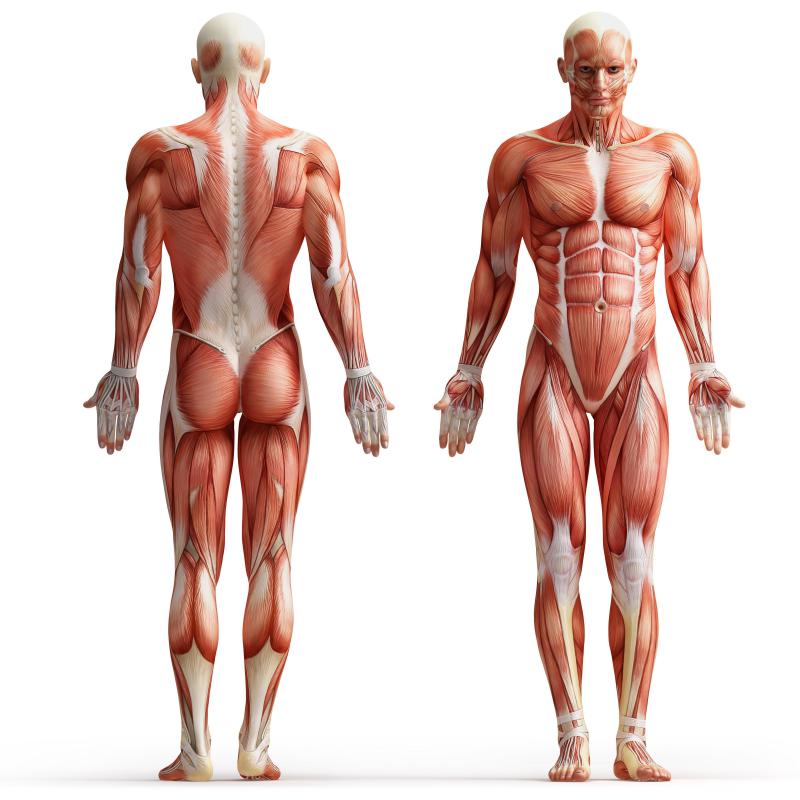
On the anterior and posterior views of the muscular system above, superficial muscles (those at the surface) are shown on the right side of the body while deep muscles (those underneath the superficial muscles) are shown on the left half of the body. For the legs, superficial muscles are shown in the anterior view while the posterior view shows both superficial and deep muscles.
Main articles: Muscular system and List of muscles of the human body
The muscular system consists of all the muscles present in a single body. There are approximately 650 skeletal muscles in the human body,but an exact number is difficult to define. The difficulty lies partly in the fact that different sources group the muscles differently and partly in that some muscles, such as palmaris longus, are not always present.The muscular system is one component of the musculoskeletal system, which includes not only the muscles but also the bones, joints, tendons, and other structures that permit movement.
Main article: muscle contractionThe three types of muscle (skeletal, cardiac and smooth) have significant differences. However, all three use the movement of actin against myosin to create contraction. In skeletal muscle, contraction is stimulated by electrical impulses transmitted by the nerves, the motoneurons (motor nerves) in particular. Cardiac and smooth muscle contractions are stimulated by internal pacemaker cells which regularly contract, and propagate contractions to other muscle cells they are in contact with. All skeletal muscle and many smooth muscle contractions are facilitated by the neurotransmitter acetylcholine.
When a sarcomere contracts, the Z lines move closer together, and the I band becomes smaller. The A band stays the same width. At full contraction, the thin and thick filaments overlap.
The action a muscle generates is determined by the origin and insertion locations. The cross-sectional area of a muscle (rather than volume or length) determines the amount of force it can generate by defining the number of sarcomeres which can operate in parallel.[citation needed] The amount of force applied to the external environment is determined by lever mechanics, specifically the ratio of in-lever to out-lever. For example, moving the insertion point of the biceps more distally on the radius (farther from the joint of rotation) would increase the force generated during flexion (and, as a result, the maximum weight lifted in this movement), but decrease the maximum speed of flexion. Moving the insertion point proximally (closer to the joint of rotation) would result in decreased force but increased velocity. This can be most easily seen by comparing the limb of a mole to a horse - in the former, the insertion point is positioned to maximize force (for digging), while in the latter, the insertion point is positioned to maximize speed (for running).
(a) Some ATP is stored in a resting muscle. As contraction starts, it is used up in seconds. More ATP is generated from creatine phosphate for about 15 seconds. (b) Each glucose molecule produces two ATP and two molecules of pyruvic acid, which can be used in aerobic respiration or converted to lactic acid. If oxygen is not available, pyruvic acid is converted to lactic acid, which may contribute to muscle fatigue. This occurs during strenuous exercise when high amounts of energy are needed but oxygen cannot be sufficiently delivered to muscle. (c) Aerobic respiration is the breakdown of glucose in the presence of oxygen (O2) to produce carbon dioxide, water, and ATP. Approximately 95 percent of the ATP required for resting or moderately active muscles is provided by aerobic respiration, which takes place in mitochondria.
Muscular activity accounts for much of the body's energy consumption. All muscle cells produce adenosine triphosphate (ATP) molecules which are used to power the movement of the myosin heads. Muscles have a short-term store of energy in the form of creatine phosphate which is generated from ATP and can regenerate ATP when needed with creatine kinase. Muscles also keep a storage form of glucose in the form of glycogen. Glycogen can be rapidly converted to glucose when energy is required for sustained, powerful contractions. Within the voluntary skeletal muscles, the glucose molecule can be metabolized anaerobically in a process called glycolysis which produces two ATP and two lactic acid molecules in the process (note that in aerobic conditions, lactate is not formed; instead pyruvate is formed and transmitted through the citric acid cycle). Muscle cells also contain globules of fat, which are used for energy during aerobic exercise. The aerobic energy systems take longer to produce the ATP and reach peak efficiency, and requires many more biochemical steps, but produces significantly more ATP than anaerobic glycolysis. Cardiac muscle on the other hand, can readily consume any of the three macronutrients (protein, glucose and fat) aerobically without a 'warm up' period and always extracts the maximum ATP yield from any molecule involved. The heart, liver and red blood cells will also consume lactic acid produced and excreted by skeletal muscles during exercise.Simplified schema of basic nervous system function. Signals are picked up by sensory receptors and sent to the spinal cord and brain via the afferent leg of the peripheral nervous system, whereupon processing occurs that results in signals sent back to the spinal cord and then out to motor neurons via the efferent leg.Efferent legThe efferent leg of the peripheral nervous system is responsible for conveying commands to the muscles and glands, and is ultimately responsible for voluntary movement. Nerves move muscles in response to voluntary and autonomic (involuntary) signals from the brain. Deep muscles, superficial muscles, muscles of the face and internal muscles all correspond with dedicated regions in the primary motor cortex of the brain, directly anterior to the central sulcus that divides the frontal and parietal lobes.In addition, muscles react to reflexive nerve stimuli that do not always send signals all the way to the brain. In this case, the signal from the afferent fiber does not reach the brain, but produces the reflexive movement by direct connections with the efferent nerves in the spine. However, the majority of muscle activity is volitional, and the result of complex interactions between various areas of the brain.
Nerves that control skeletal muscles in mammals correspond with neuron groups along the primary motor cortex of the brain's cerebral cortex. Commands are routed though the basal ganglia and are modified by input from the cerebellum before being relayed through the pyramidal tract to the spinal cord and from there to the motor end plate at the muscles. Along the way, feedback, such as that of the extrapyramidal system contribute signals to influence muscle tone and response.Deeper muscles such as those involved in posture often are controlled from nuclei in the brain stem and basal ganglia.Afferent leg
The afferent leg of the peripheral nervous system is responsible for conveying sensory information to the brain, primarily from the sense organs like the skin. In the muscles, the muscle spindles convey information about the degree of muscle length and stretch to the central nervous system to assist in maintaining posture and joint position. The sense of where our bodies are in space is called proprioception, the perception of body awareness. More easily demonstrated than explained, proprioception is the "unconscious" awareness of where the various regions of the body are located at any one time. This can be demonstrated by anyone closing their eyes and waving their hand around. Assuming proper proprioceptive function, at no time will the person lose awareness of where the hand actually is, even though it is not being detected by any of the other sensesSeveral areas in the brain coordinate movement and position with the feedback information gained from proprioception. The cerebellum and red nucleus in particular continuously sample position against movement and make minor corrections to assure smooth motion.There is also a companion magazine called Muscle and Fitness Hers oriented toward women.
Muscle & Fitness Fitness Exercise for Women for Men for Women at Home for Men at Home Abs For Kids for Women to Lose Weight Tumblr Photos

Muscle & Fitness Fitness Exercise for Women for Men for Women at Home for Men at Home Abs For Kids for Women to Lose Weight Tumblr Photos
Muscle & Fitness Fitness Exercise for Women for Men for Women at Home for Men at Home Abs For Kids for Women to Lose Weight Tumblr Photos

Muscle & Fitness Fitness Exercise for Women for Men for Women at Home for Men at Home Abs For Kids for Women to Lose Weight Tumblr Photos
Muscle & Fitness Fitness Exercise for Women for Men for Women at Home for Men at Home Abs For Kids for Women to Lose Weight Tumblr Photos

Muscle & Fitness Fitness Exercise for Women for Men for Women at Home for Men at Home Abs For Kids for Women to Lose Weight Tumblr Photos

Muscle & Fitness Fitness Exercise for Women for Men for Women at Home for Men at Home Abs For Kids for Women to Lose Weight Tumblr Photos
 Muscle & Fitness Fitness Exercise for Women for Men for Women at Home for Men at Home Abs For Kids for Women to Lose Weight Tumblr Photos
 Muscle & Fitness Fitness Exercise for Women for Men for Women at Home for Men at Home Abs For Kids for Women to Lose Weight Tumblr Photos
 Muscle & Fitness Fitness Exercise for Women for Men for Women at Home for Men at Home Abs For Kids for Women to Lose Weight Tumblr Photos
 Muscle & Fitness Fitness Exercise for Women for Men for Women at Home for Men at Home Abs For Kids for Women to Lose Weight Tumblr Photos
No comments:
Post a Comment